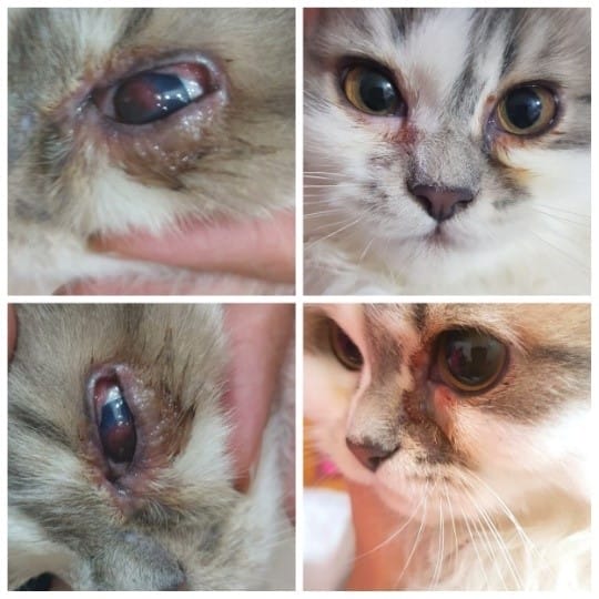
Keratoconjunctivitis sicca
Keratoconjunctivitis sicca KCS
Keratoconjunctivitis sicca, the fancy way to say the eye is dry, usually arises from inadequate production of watery tears, The tear film is a crucial component of cat eye health it’s normally consists of Lipids, Aqueous fluid and mucin which lubricate the eye and act as a barrier protecting it from dehydration or invasion by microorganisms.
:Predisposed Breeds to Keratoconjunctivitis sicca KCS
West Highland white terrier, Pug, English cocker spaniel, English springer, spaniel, English bulldog, Toy poodle.
The highest occurrence of the disease was observed at the age between 4 and 7 years.
:Causes of KCS
1- Immune-mediated inflammation of the tear gland
2-Feline Herpes Virus Infection
3-Trauma
4-Corneal Ulceration
5-Damage to facial nerve
التهاب القرنية والملتحمة المصحوب بجفاف العين نتيجة لقلة افراز الدموع المرطبة للعين.
كثيراً ما تكون امراض العيون في القطط لعدة أسباب منها، الأمراض الفيروسية في المرتبة الأولي و عادةً تصيب هذه العدوي الفيروسية القطط التي لم تحصل علي التطعيمات الوقائية بجدول زمني سليم او لم تأخذها بالأساس. لذلك وجب التنويه عن أهمية التطعيمات الوقائية صد الأمراض الفيروسية بداية من عمر 45 يوماً و حتي عمر 4 أشهر بمعدل جرعة كل 3-4 أسابيع.
:Case history
Owned 3 y old cat with unclear vaccination profile represented with asymmetry between both eyes, swelled eyelids inflamed eyelids and conjunctival membrane, ocular yellow to gray discharges, cloudy dull eye, blepharospasms and photosensitivity.
:Clinical Signs
![]() Blepharospasms: Is frequently observed as a first sign of the disease. It is caused by corneal irritation as a result of changes in the tear fluid. It may be associated with photophobia.
Blepharospasms: Is frequently observed as a first sign of the disease. It is caused by corneal irritation as a result of changes in the tear fluid. It may be associated with photophobia.
![]() Mucoid or mucopurulent discharge: In the absence of the aqueous layer of the tear film, mucoid layer of the tear film is insufficiently eliminated from the eye and can be seen dry around the palpebral rim. This is together with conjunctivitis one of the early signs of KCS.
Mucoid or mucopurulent discharge: In the absence of the aqueous layer of the tear film, mucoid layer of the tear film is insufficiently eliminated from the eye and can be seen dry around the palpebral rim. This is together with conjunctivitis one of the early signs of KCS.
![]() Corneal ulceration: Is described mainly in chronic cases where loss of epithelium occurs in the central corneal area. This condition may lead to corneal perforation and endophthalmitis.
Corneal ulceration: Is described mainly in chronic cases where loss of epithelium occurs in the central corneal area. This condition may lead to corneal perforation and endophthalmitis.
![]() Corneal vascularization and pigmentation: Deepness and extent of the corneal changes correlates with the disease chronicity.
Corneal vascularization and pigmentation: Deepness and extent of the corneal changes correlates with the disease chronicity.
![]() Corneal xerosis and conjunctival redness: Dry appearance of the cornea is typical.
Corneal xerosis and conjunctival redness: Dry appearance of the cornea is typical.
![]() Dry ipsilateral nostril.
Dry ipsilateral nostril.
![]() Chronic staphylococcus infection with good responses to antibiotics.
Chronic staphylococcus infection with good responses to antibiotics.
:Ophthalmic examination
–Ocular examination showed hyperemic conjunctiva with tortuous scleral blood vessels, mucopurulent discharges, yellow crusts sticking on eye corners, opaque corneal.
-With Schirmer tear test special paper inserted in the lower eyelid in the outer corner of the eye, the moisten paper height reached 10mm which was indication for eye dryness.
-Fluorescein stain was negative for corneal ulceration.
.Diagnosis directed to Keratoconjunctivitis sicca.
Eye cytology, culture and sensitivity was recommended characterizing bacterial infection and appropriate antibiotic ttt.

:Treatment plan
:Treatment plan for Keratoconjunctivitis sicca included in
. Artificial tears and TBH it won’t replace the previously secreted tear film and it lacks mucins and lipids but it’s help in the eye lubrication
.Dexpanthenol gel as eye lubricant
Stem cells therapy: Mesenchymal stem cells (MSCs) applied topically on the ocular surface treating the dry eye condition as regenerative therapy.
Platelets rich plasma QID: Prepared by drawing blood sample from the patient separating the plasma and applying it topically on the ocular surface.
.Moxifloxacin as Ocular broad spectrum antibiotic eye drop
.Topical NSAIDs eye drops after excluding corneal ulcerations
Improvement seen after 3wks after initiating treatment plan Alhamdullah.
Thanks to the teamwork for continually raising the level of performance and achievement in of keratoconjunctivitis sicca case, and specially for Dr. Ahmed Barakat for his dedication in this case dealing with such destroyed eye and initiating amazing line of treatment in Egyptian veterinary field at this time like stem cells.

Dr.Ahmad Barakat
Postgraduate degree holder in Small animal soft tissue surgeries. Published more than 10 medical articles. A certified lecturer in . Small animal soft tissue surgery . Lieutenant at Egyptian army for 3 year . specialist in orthopedic surgery and cclr from 2016. Medical Consultant in pharmaceutical company . on of the pioneers in Advanced radiological diagnostics imaging as CT, MRI . Oncological clinical management and analysis. . First to contribute in minimal invasive surgery and endoscopy .
